Uncategorized files
Jump to navigation
Jump to search
Showing below up to 50 results in range #1 to #50.
-
 14 and 6 Positive Bursts.png 1,033 × 631; 240 KB
-
 6 Hertz Phantom Spike Wave Discharges.png 1,039 × 646; 231 KB
-
 7-month-old posterior dominant rhythm.png 1,384 × 830; 875 KB
-
 7 Hz posterior rhythm.png 1,318 × 794; 772 KB
-
 8 year old EEG.png 1,110 × 666; 570 KB
-
 95 year old.png 1,136 × 696; 658 KB
-
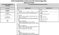 ACNS Reference Chart.png 1,161 × 684; 275 KB
-
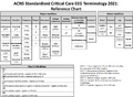 ACNS Reference Chart001.png 1,038 × 763; 340 KB
-
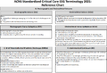 ACNS Reference Chart002.png 1,019 × 698; 383 KB
-
 ACNS Standardized Critical Care EEG Terminology 2021- Reference Chart.png 1,055 × 680; 300 KB
-
 A 60 Hz notch filter.png 1,083 × 572; 178 KB
-
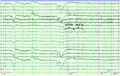 Alpha Squeak.png 688 × 434; 309 KB
-
 Anterior-posterior voltage and frequency gradients.png 826 × 654; 123 KB
-
 Anterior posterior longitudinal bipolar montage.png 1,600 × 1,083; 584 KB
-
 Appearance of hypnagogic hypersynchrony and triangular vertex waves.png 1,348 × 938; 744 KB
-
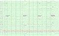 Artifacts001.png 721 × 446; 192 KB
-
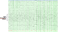 Breach rhythm.png 1,078 × 598; 254 KB
-
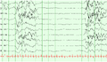 Burst Suppression Pattern.png 623 × 366; 73 KB
-
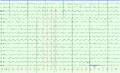 Cardioballistogram.png 1,028 × 627; 177 KB
-
 Central-Midline Theta Rhythm of Ciganek.png 847 × 649; 287 KB
-
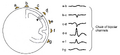 Chain of bipolar channels.png 1,650 × 752; 158 KB
-
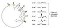 Chain of referential channels.png 1,592 × 776; 159 KB
-
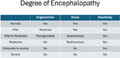 Death of Encephalopathy.png 459 × 220; 39 KB
-
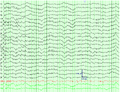 Dominant Frequency001.png 861 × 657; 292 KB
-
 Dominant Frequency is in the alpha range with loss of reactivity.png 1,041 × 626; 284 KB
-
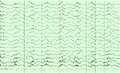 Dominant frequency002.png 1,038 × 629; 254 KB
-
 EEG Analysis of Waveforms - Frequency.png 2,234 × 328; 55 KB
-
 EEG Analysis of Waveforms - Morphology.png 2,202 × 290; 87 KB
-
 EEGmasterLogo.png 454 × 350; 147 KB
-
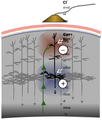 Electrical-Principles.png 744 × 876; 119 KB
-
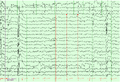 Electrocardiographic Artifacts.png 896 × 613; 265 KB
-
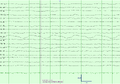 Electrocerebral Inactivity (ECI).png 995 × 696; 137 KB
-
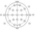 Electrode-placement.jpg 270 × 246; 16 KB
-
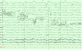 Electrode pops.png 1,041 × 629; 226 KB
-
 Electromyographic Artifacts.png 1,042 × 626; 191 KB
-
 Electroretinogram.png 979 × 598; 228 KB
-
 Fast alpha variant.png 853 × 521; 210 KB
-
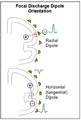 Focal Discharge Dipole Orientation.png 356 × 526; 42 KB
-
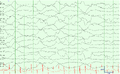 Galvanic Skin Responses.png 1,040 × 638; 209 KB
-
 Generalized Periodic Discharges with Triphasic Morphology.png 1,044 × 691; 227 KB
-
 Generalized Rhythmic Delta Activity (GRDA) frontally predominant.png 942 × 660; 271 KB
-
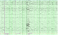 Glossokinetic Artifacts.png 1,047 × 624; 314 KB
-
 Image.png 721 × 592; 348 KB
-
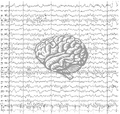 Image001 introduction-to-eeg.png 938 × 890; 369 KB
-
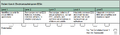 Image002 neurology-residency-milestones.png 1,835 × 537; 125 KB
-
 Image004 first-recorded-eeg.jpg 300 × 61; 6 KB
-
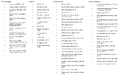 Images003 must-know.png 1,250 × 781; 288 KB
-
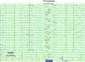 K-complexes during N2 Sleep.png 760 × 557; 187 KB
-
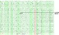 Lambda waves.png 1,034 × 618; 248 KB
-
 Logo transp eegmaster.png 521 × 447; 110 KB


.png)


_frontally_predominant.png)


